KVI-CART researchers aim for real-time proton therapy
28 February 2019
The researchers aim to visualize short-lived positron emitter isotopes, during proton radiotherapy treatment.
Proton therapy is a highly accurate irradiation technique. The proton beam used for treatment can be steered towards (every part of) the tumor, minimizing radiation damage to surrounding healthy tissues. Prior to the treatment, the direction and depth of penetration of the proton beam are accurately determined using acquired (CT) images, and validated by means of quality assurance measurements. But it is even better to (also) measure treatment accuracy within the patient.
Positron-emission tomography (PET), which measures gamma radiation released after protons and atomic nuclei collide, allows for such a measurement. Several groups worldwide investigate and apply this methodology, using so-called long-lived positron emitters. This has two major drawbacks. First, acquiring the PET-images takes a substantial amount of time allowing image acquisition only after completion of the radiation treatment. A second drawback is that the time elapsed between the start of radiation and end of PET-image acquisition allows these long-lived positron emitters to travel through the patient body. This reduces the accuracy of the image.
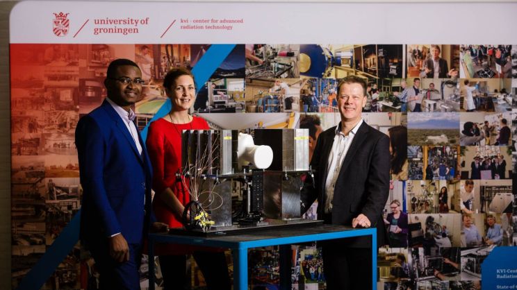
The KVI-CART researchers Peter Dendooven and Ikechi Azoemelam, in close collaboration with Siemens, investigate an alternative method using short-lived positron emitters. They are the only group worldwide having a PET-scanner that can very quickly be turned on and off. Images can therefore be acquired during the radiation treatment, allowing the real-time adaptation of the treatment, should that be indicated. The researchers currently use more and more intricate phantoms as a proof of concept. Subsequent clinical application of their new method may require another few years of product development.
For more details regarding this research, please see this article (in dutch).
 Image copyright Siemens Healthcare

 English
 Nederlands