Image biomarkers to improve prediction of side effects
Image biomarkers of CT, PET and MR imaging of parotid glands to improve prediction of xerostomia
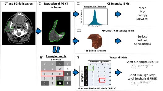
Conventional Normal Tissue Complication Probability (NTCP) models that predict patient-rated side-effects are mainly based on dose-volume parameters and baseline complaints. Using these models, however, there still is a substantial and unexplained variance in the prediction of side-effects. In order to improve the identification of patients at risk, anatomical and physiological information is extracted from diagnostic images - such as CT, 18FDG PET and MR images - and quantified in so called image biomarkers. We focus on improving the prediction of side-effects by using these image biomarkers.
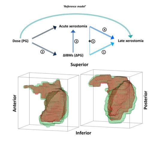
Changes in image biomarkers in CT images of parotid glands to improve the predict of xerostomia
By investigating the changes of the parotid glands during and after radiotherapy, we aim to improve the understanding of the development of radiation-induced xerostomia. By quantifying these changes, our aim is to complement the current assessment of xerostomia that uses patient or physician rated scores, or to find a surrogate.
People involved
Sanne van Dijk, Marianna Sijtsema, Roel Steenbakkers, Hans Langendijk, Charlotte Brouwer, Arjen van der Schaaf, Tian Tian Zhai.
References
van Dijk L V., Brouwer CL, van der Schaaf A, et al: CT image biomarkers to improve patient-specific prediction of radiation-induced xerostomia and sticky saliva. Radiother Oncol 2016 122:185–191 (pdf)
Dijk LV Van, Noordzij W, Brouwer CL, et al: 18F-FDG PET image biomarkers improve prediction of late radiation-induced xerostomia. Radiother Oncol. 2018 126(1):89-95 (pdf)
Dijk LV Van, Brouwer CL, et al: CT image biomarker changes of the parotid gland are associated with late xerostomia. Int J Radiat Oncol Biol Phys submitted

 English
 Nederlands

