 English
 Nederlands
Image biomarkers to improve prediction of treatment outcome
Image biomarkers of CT imaging of head and neck tumors and lymph nodes to improve the prediction of survival and local-regional control
Recent studies have demonstrated that image biomarkers are significantly associated with treatment outcome. We tested whether the performance of prediction models for local recurrence, regional recurrence, disease free survival and overall survival could be improved by the addition of image biomarkers. Using these image biomarkers, we will furthermore focus on the pre-treatment patient-individual prediction of various endpoints.
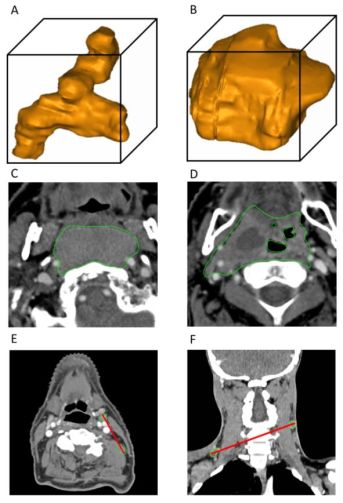 Examples of patients with low (A) and high (B) values of volume-density of the tumor.
Examples of patients with low (C) and high (D) values of Run Length Non-uniformity of the tumor. Examples of Major-axis-length in patients with two unilateral (E) and two bilateral (F) positive lymph nodes.
People involved
Tiantian Zhai, Sanne van Dijk, Marianna Sijtsema, Roel Steenbakkers, Hans Langendijk, Charlotte Brouwer, Arjen van der Schaaf.
References
T.T. Zhai et al. Improving the prediction of overall survival for head and neck cancer patients using image biomarkers in combination with clinical parameters. Radiother Oncol. 2017 124(2):256-262 (pdf)
T.T. Zhai et al. Prognostic image biomarkers for nasopharyngeal cancer patients treated with (chemo)radiation. Abstract 2918 at 2016 ASTRO.